For most of us, upon waking in the morning we sit over the edge of the bed, stretch our ribs and shoulders, amble to the bathroom, grip our toothbrush, clean our teeth, open the shower door, step into the shower, turn the mixer handle, wash our body, undo the hair shampoo bottle, wash our hair, towel dry, open the lid of the moisturiser bottle apply all over, open the shower door, dress, tie the laces of our shoes then go to the kitchen for breakfast. Nothing extraordinary in that — we take this daily rite for granted.
If we rewind and then in slow motion replay this ritual and watch what our hands have performed we may be humbled. A new robotic hand has impressed engineers around the world by being able to handle tweezers, pour a drink, crush a beer can, and gently grip an egg. However, all the brain power of our best scientists have yet to create a robotic hand that could even crudely move us from bed to bath and beyond. We must pay more attention to our hand health.
The human hand is made up of the wrist, palm, and fingers, and consists of 27 bones, 27 joints, 34 muscles, over 100 ligaments and tendons, and many blood vessels and nerves.
The hands enable us to perform many of our daily activities such as driving, writing and cooking. It is important to understand the normal anatomy of the hand to learn more about diseases and conditions that can affect them.
A quick lesson in hand anatomy
Motion Orthopaedics has created a very good YouTube visual display if you want to understand more. www.motionorthodocs.com/hand-anatomy-ortho-surgeon-creve-coeur-saint-louis.html
Bones
The wrist is comprised of eight carpal bones. These wrist bones are attached to the radius and ulna of the forearm to form the wrist joint. They connect to five metacarpal bones that form the palm of the hand. Each metacarpal bone connects to one finger at a joint called the metacarpophalangeal joint or MCP joint. The knuckle joint as you may know it.
The bones in our fingers and thumb are called phalanges. Each finger has 3 phalanges separated by two interphalangeal joints, except for the thumb, which only has 2 phalanges and one interphalangeal joint.
The first joint close to the knuckle is called the proximal interphalangeal joint or PIP joint. The joint closest to the end of the finger is called the distal interphalangeal joint or DIP joint.
The MCP joint and the PIP joint act like hinges when the fingers bend and straighten.
Soft tissues
Our hand bones are held in place and supported by various soft tissues. These include: articular cartilage, ligaments, muscles, and tendons.
Articular cartilage is a smooth material that acts as a shock absorber and cushions the ends of bones at each of the 27 joints, allowing smooth movement of the hand.
Muscles, tendons, and ligaments function to control the movement of the hand.
Ligaments are tough rope-like tissue that connect bones to other bones, holding them in place and providing stability to the joints. Each finger joint has two collateral ligaments on either side, which prevents the abnormal sideways bending of the joints. The volar plate is the strongest ligament in the hand. It joins the proximal and middle phalanx on the palm side of the joint and prevents backwards bending of the PIP joint (hyperextension).
Muscles
Muscles are fibrous tissues that help produce movement. Muscles work by contracting.
There are two types of muscles in the hand, intrinsic and extrinsic muscles.
Intrinsic muscles are small muscles that originate in the wrist and hand. They are responsible for fine motor movement of the fingers during activities such as writing or playing the piano.
Extrinsic muscles originate in the forearm or elbow and control the movement of the wrist and hand. These muscles are responsible for gross hand movements. They position the wrist and hand while the fingers perform fine motor movements.
Each finger has six muscles controlling its movement: three extrinsic and three intrinsic muscles. The index and little finger each have an extra extrinsic extensor.
Tendons
Tendons are soft tissues that connect muscles to bones. When muscles contract, tendons pull the bones causing the finger to move. The extrinsic muscles attach to finger bones through long tendons that extend from the forearm through the wrist. Tendons located on the palm side help in bending the fingers and are called flexor tendons, while tendons on top of the hand help in straightening the fingers, and are called extensor tendons.
Nerves
Nerves of the hand carry electrical signals from the brain to the muscles in the forearm and hand, enabling movement. They also carry the senses of touch, pain, and temperature back from the hands to the brain.
The three main nerves of the hand and wrist are the ulnar nerve, radial nerve and median nerve. All three nerves originate at the shoulder and travel down the arm to the hand. Each of these nerves has sensory and motor components.
Ulnar nerve: The ulnar nerve crosses the wrist through an area called Guyon’s canal and branches to provide sensation to the little finger and half of the ring finger.
Median nerve: The median nerve crosses the wrist through a tunnel called the carpal tunnel. The median nerve provides sensation to the palm, thumb, index finger, middle finger, and part of the ring finger.
Radial nerve: The radial nerve runs down the thumb side of the forearm and provides sensation to the back of the hand from the thumb to the middle finger.
Blood vessels
Blood vessels travel beside the nerves to supply blood to the hand. The main arteries are the ulnar and radial arteries, which supply blood to the front of the hand, fingers, and thumb.
The ulnar artery travels next to the ulnar nerve through the Guyon’s canal in the wrist.
The radial artery is the largest artery of the hand, traveling across the front of the wrist, near the thumb. Pulse is measured at the radial artery.
Other blood vessels travel across the back of the wrist to supply blood to the back of the hand, fingers, and thumb.
Bursae
Bursae are small fluid filled sacs that decrease friction between tendons and bone or skin. Bursae contain special cells called synovial cells that secrete a lubricating fluid.
Hand health
While the hand is complex, and there is medical delight in asking a patient “How fit is your extensor carpi radialis longus today?” we’ll keep the hand fitness programme simple, but very effective for the busy judge. Here are some exercises for you to try.
Exercise one: prayer stretch
Put hands together, forward facing, spread elbows but be sure to not lose hand contact. Slowly turn hands to prayer position, hold 10–20 seconds, repeat twice.
Exercise two: desk stretch
Place flats of hands on desk. Make sure all fingers are on the desk or table. Lean back. Hold 10–20 seconds, repeat twice. Alleviates writer’s cramp or keyboard hand fatigue.
Exercise three: strengthen fingers, hands, and wrists
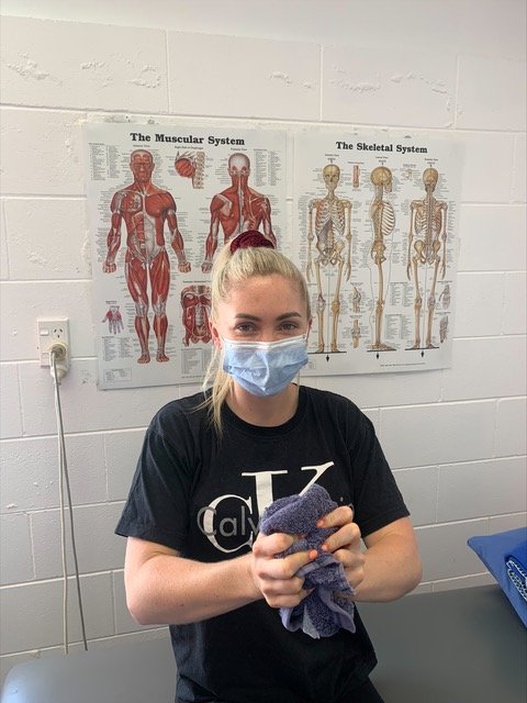
Bunch a small towel, squeeze, twist, pull. Do this for five minutes in front of television, four to five times per week. This will make a substantial change in finger and hand strength.
A happy and healthy new year to you all. Keep fit.
Rebecca Mooney & Malcolm Hood


